Anatomy Of Chest ~ Human Chest Anatomy Illustration Stock Image F025 1029 Science Photo Library. It also protects several vital organs of the chest, such as the heart, aorta, vena cava, and. For example, you wake up at night with intense stomach pain. Several muscles that move the arms, head, and neck have their origins on the sternum. Here, we break down the anatomy of your chest muscles. Thoracic cavity, also called chest cavity, the second largest hollow space of the body.
Browse 6,407 chest anatomy stock photos and images available, or search for human anatomy to find more great stock photos and pictures. Anatomy is the science that studies the structure of the body. Plus, how to target each to make them bigger and stronger. The upper torso is considered to be anything above the waist and below the neck, including the shoulders and back. Here's how science can help you grow!
Male Anatomy Chest Anatomy Drawing Diagram from t3.ftcdn.net Anatomy is the science that studies the structure of the body. 30 lines of the thoracic wall syllabus p. Sternocleidomastoid muscle clavicle and ribs anatomy muscle anatomy chest sternocleidomastoid ribs anatomy chest muscles anatomy thorax rib muscles chest muscles chest anatomy illustration. See chest anatomy stock video clips. Thoracic cavity, also called chest cavity, the second largest hollow space of the body. It is important to remember the position and orientation of the heart when placing a stethoscope on the chest of a patient and listening for heart sounds, and also when looking at images taken from a midsagittal perspective. Use the mouse scroll wheel to move the images up and down alternatively use the tiny arrows (>>) on both side of the image to move the images.>>) on both side of the image to move the images. This thoracic and pulmonary anatomy tool is especially designed for students of anatomy (medical and paramedical studies).
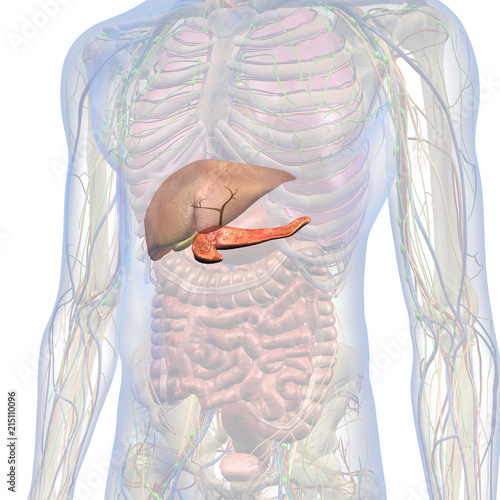
The chest is the area of origin for many of the body's systems as it houses organs such as the heart, esophagus, trachea, lungs, and thoracic diaphragm.
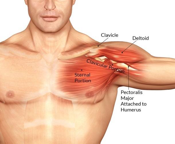
Thoracic cavity, also called chest cavity, the second largest hollow space of the body. 31 anatomy of the female breast syllabus p. For example, you wake up at night with intense stomach pain. About the 6th week, the somites differentiate into the sclerotomes and the dermatomyotomes. See human chest anatomy stock video clips. Muscular anatomy of the chest pectoralis major. Several muscles that move the arms, head, and neck have their origins on the sternum. Anatomy is the science that studies the structure of the body. The right side of the heart is deflected anteriorly, and the left side is deflected posteriorly. This page provides an overview of the chest muscle group. The upper torso is considered to be anything above the waist and below the neck, including the shoulders and back. Get the full built by science program: The pectoralis major and the pectoralis minor, known collectively as your pecs.
The sternum, commonly known as the breastbone, is a long, narrow flat bone that serves as the keystone of the rib cage and stabilizes the thoracic skeleton. How to view the anatomical labels. Chest a man's chest — like the rest of his body — is covered with skin that has two layers. Several muscles that move the arms, head, and neck have their origins on the sternum. Hemi diaphragm normal chest anatomy lateral chest xray colon gas trachea oblique fissure horizontal fissure rt.
Chest Muscles Anatomy Bodybuilding Wizard from bodybuilding-wizard.com Sternocleidomastoid muscle clavicle and ribs anatomy muscle anatomy chest sternocleidomastoid ribs anatomy chest muscles anatomy thorax rib muscles chest muscles chest anatomy illustration. It provides access to ct images in the axial plane, allowing the user to learn and review the lung anatomy interactively. System respiratory respiratory organs of human body digestive and respiratory system medical chest internal structure of human body medicine body lungs biology intestines stomach anatomy torso human internal. It also protects several vital organs of the chest, such as the heart, aorta, vena cava, and. A good radiologist knows the anatomy because knowing where structures normally live and recognizing the location of an abnormality helps to make or narrow the differential diagnosis. This page provides an overview of the chest muscle group. Chest a man's chest — like the rest of his body — is covered with skin that has two layers. The chest is made up primarily of two muscles:
Chest a man's chest — like the rest of his body — is covered with skin that has two layers.
Here, we break down the anatomy of your chest muscles. The chest is the area of origin for many of the body's systems as it houses organs such as the heart, esophagus, trachea, lungs, and thoracic diaphragm. The pectoralis major and the pectoralis minor, known collectively as your pecs. Use the mouse scroll wheel to move the images up and down alternatively use the tiny arrows (>>) on both side of the image to move the images.>>) on both side of the image to move the images. Radiology basics of chest ct anatomy with annotated coronal images and scrollable axial images to help medical students and junior doctors learning anatomy. 4 innervation of the breast blood supply of the breast syllabus p. A detailed knowledge of chest wall anatomy is crucial for reconstructions in this difficult patient population. See chest anatomy stock video clips. The chest is made up primarily of two muscles: 30 lines of the thoracic wall syllabus p. The chest anatomy includes the pectoralis major, pectoralis minor and the serratus anterior. It provides access to ct images in the axial plane, allowing the user to learn and review the lung anatomy interactively. 31 anatomy of the female breast syllabus p.
The myotomes elongate and invade the mesoderm of the wall of the embryonic thoracic and abdominal cavities. A detailed knowledge of chest wall anatomy is crucial for reconstructions in this difficult patient population. Anatomy of the chest and the lungs: Radiology basics of chest ct anatomy with annotated coronal images and scrollable axial images to help medical students and junior doctors learning anatomy. Download my two educational text books for free using this link:
Anatomy For Artists Chest Abs Youtube from i.ytimg.com These myotomes divide into the epimere and the hypomere. In insects, crustaceans, and the extinct trilobites, the thorax is one of the three main divisions of the creature's body, each of which is in turn composed of multiple segments. About the 6th week, the somites differentiate into the sclerotomes and the dermatomyotomes. The muscles of the chest develop from the somites found in the mesoderm. (1) the pectoralis major, and (2) the pectoralis minor. The chest or thorax is the region between the neck and diaphragm that encloses organs, such as the heart, lungs, esophagus, trachea, and thoracic diaphragm. The chest is the area of origin for many of the body's systems as it houses organs such as the heart, esophagus, trachea, lungs, and thoracic diaphragm. This chapter is an abbreviated review of thoracic anatomy as seen on chest radiographs and computed tomography (ct) of the chest.
For example, you wake up at night with intense stomach pain.
Anatomy is the science that studies the structure of the body. The circulatory system does most of its work. It spreads out like a fan and covers the rib cage like an armor plate. For example, you wake up at night with intense stomach pain. Get the full built by science program: Chest a man's chest — like the rest of his body — is covered with skin that has two layers. Plus, how to target each to make them bigger and stronger. System respiratory respiratory organs of human body digestive and respiratory system medical chest internal structure of human body medicine body lungs biology intestines stomach anatomy torso human internal. Chest muscles anatomy (1) pectoralis major muscle. Fill out your shirt with a bigger, stronger, more powerful chest. A part of the upper torso, the chest is the are. Learn about each of these muscles, their locations, functional anatomy and exercises for them. It provides protection to vital organs (eg, heart and major vessels, lungs, liver) and provides stability for movement.